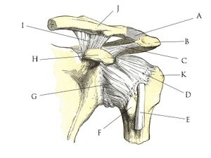
Conjoint Tendon Shoulder Anatomy - Dynamic And Static Stabilization Of Anterior Shoulder Instability With The Subscapular Sling Procedure Arthroscopy Techniques : The conjoint tendon formed by the short head of biceps brachii and coracobrachial muscles is attached to the tip of the cp.
byAdmin-
0
Conjoint Tendon Shoulder Anatomy - Dynamic And Static Stabilization Of Anterior Shoulder Instability With The Subscapular Sling Procedure Arthroscopy Techniques : The conjoint tendon formed by the short head of biceps brachii and coracobrachial muscles is attached to the tip of the cp.. Subscapularis arises, as the name suggests, from the undersurface of the scapula and is an internal rotator of the shoulder. Shoulder muscles and shoulder tendons. Becomes lateral antebrachial cutaneous nerve (terminal branch) emerges lateral to distal bicep tendon and brachoradialis to form lateral antebrachial cutaneous nerve. Thus, the biceps muscle, which functions to bend the elbow and rotate the forearm, has two anchor points in the shoulder region. Anatomy textbooks describe separate insertion sites for these two tendons.
Subscapularis arises, as the name suggests, from the undersurface of the scapula and is an internal rotator of the shoulder. Becomes lateral antebrachial cutaneous nerve (terminal branch) emerges lateral to distal bicep tendon and brachoradialis to form lateral antebrachial cutaneous nerve. Conjoint tendon tenotomy tenotomy of the conjoint tendon is performed from within the glenohumeral joint on the posterior aspect of the subscapularis where the coracoid graft and hardware make contact with the subscapularis muscle medially. Other important bones in the shoulder include: Anatomy, abdomen and pelvis, conjoint tendon (inguinal aponeurotic falx).
Conjoint Tendon from hyperleap.com The short head of the bicep is a continuation of the conjoined tendon, which originates from a bony hook (the coracoid) at the front of the shoulder blade. Iatrogenic injury of the axillary and subscapular nerves: The glenohumeral joint is an inherently unstable joint and depends on the surrounding soft tissues for stabilization. It forms the medial part of the posterior wall of the inguinal canal. Do not extend dissection medial to the glenoid. The acromion is a bony. Locate a nonabsorbable suture from the initial surgery for orientation. Teres minor conjoint tendon •anterior •superior.
The conjoint tendon then turns inferiorly and attaches onto the pubic crest and pecten pubis 1.
The glenohumeral joint is an inherently unstable joint and depends on the surrounding soft tissues for stabilization. Follow muscle fibers of the conjoint tendon superiorly to locate the coracoid remnant. The coracobrachialis and short head of biceps originate from the coracoid and inserts separately into the anterior humerus and the biciptal tuberosity of the ulna and lacertus fibrosis of the forearm. Thus, the biceps muscle, which functions to bend the elbow and rotate the forearm, has two anchor points in the shoulder region. Iatrogenic injury of the axillary and subscapular nerves: Cal, cp and the conjoint tendon should be evaluated as an important. The fascia on the lateral side of the conjoint tendon is incised to reveal the subscapularis external rotation puts the subscapularis fibers on stretch Asheesh bedi md, in shoulder and elbow injuries in athletes, 2018. Shoulder muscles and shoulder tendons. Other important bones in the shoulder include: It is a multipennate muscle forming several tendons that insert as a conjoined unit on the medial border of the bicipital groove. During a surgical procedure, which included releasing these two tendons off the humerus from a posterior approach, they appeared to be joined together. • under normal conditions the amount of friction is reduced to a minimum by the large subacromial bursa, which shoulder tendon anatomy.
Cadaver shoulders were subsequently dissected to determine if the tendons had conjoint or separate insertions. Shoulder muscles and shoulder tendons. Shoulder radiology & anatomy at usuhs.mil. The short head of the bicep is a continuation of the conjoined tendon, which originates from a bony hook (the coracoid) at the front of the shoulder blade. Teres minor conjoint tendon •anterior •superior.
Biomechanics Of The Shoulder Physiopedia from www.physio-pedia.com The coracobrachialis and short head of biceps originate from the coracoid and inserts separately into the anterior humerus and the biciptal tuberosity of the ulna and lacertus fibrosis of the forearm. Becomes lateral antebrachial cutaneous nerve (terminal branch) emerges lateral to distal bicep tendon and brachoradialis to form lateral antebrachial cutaneous nerve. Thus, the biceps muscle, which functions to bend the elbow and rotate the forearm, has two anchor points in the shoulder region. Pointing laterally forward, it, together with the acromion, serves to stabilize the shoulder joint. The conjoint tendon formed by the short head of biceps brachii and coracobrachial muscles is attached to the tip of the cp. Asheesh bedi md, in shoulder and elbow injuries in athletes, 2018. Conjoint tendon tenotomy for glenoid exposure in the setting of. Bristow procedure is performed when there is bone loss in the front of glenoid cavity with multiple dislocations of the shoulder and the coracoid is transferred to that area after osteotomy.
Do not extend dissection medial to the glenoid.
Conjoint tendon shoulder anatomy conjoint tendon shoulder anatomy / abdomen | basicmedical key. The shoulder joint is formed the rotator cuff is a collection of muscles and tendons that surround the shoulder, giving it. Iatrogenic injury of the axillary and subscapular nerves: Cadaver shoulders were subsequently dissected to determine if the tendons had conjoint or separate insertions. The tendons of insertion of the latissimus dorsi and the teres major muscles and the tendon of origin of the long head of the triceps brachii muscle were united, forming a conjoint tendon that attached to the infraglenoid tubercle of the scapula and the lower part of the anatomical neck of the humerus adhering to the articular capsule of the. Shoulder radiology & anatomy at usuhs.mil. Locate a nonabsorbable suture from the initial surgery for orientation. Retraction of the conjoint tendon must be done with care. Do not extend dissection medial to the glenoid. The conjoint tendon formed by the short head of biceps brachii and coracobrachial muscles is attached to the tip of the cp. Asheesh bedi md, in shoulder and elbow injuries in athletes, 2018. Anatomy of the axillary nerve and its relation to inferior capsular shift. • under normal conditions the amount of friction is reduced to a minimum by the large subacromial bursa, which shoulder tendon anatomy.
(1)royal columbian hospital, new westminster, canada. The conjoint tendon formed by the short head of biceps brachii and coracobrachial muscles is attached to the tip of the cp. Teres minor conjoint tendon •anterior •superior. The conjoint tendon then turns inferiorly and attaches onto the pubic crest and pecten pubis 1. • under normal conditions the amount of friction is reduced to a minimum by the large subacromial bursa, which shoulder tendon anatomy.
Ki Jinn Chin On Twitter These Images Are What I Base The Term Conjoint Tendon On Although I Appreciate The Structure Seen May Not Actually Arise From Teres Major Or Lat Dorsi from pbs.twimg.com The conjoint tendon then turns inferiorly and attaches onto the pubic crest and pecten pubis 1. It is a multipennate muscle forming several tendons that insert as a conjoined unit on the medial border of the bicipital groove. Dissection of the rotator interval: With retraction of deltoid, the distal section of the pectoralis major, which inserts on the crest of the greater tubercle is visualised. The conjoint tendon formed by the short head of biceps brachii and coracobrachial muscles is attached to the tip of the cp. It forms the medial part of the posterior wall of the inguinal canal. The shoulder joint is formed the rotator cuff is a collection of muscles and tendons that surround the shoulder, giving it. The muscles form a conjoint tendon and flex the shoulder as well as the elbow.
The tendon measures approximately 8 cm from superior to inferior.
Shoulder anatomy for ultrasound evaluation. A detailed understanding of the rotator cuff anatomy and its associated structures is required of the treating physician. Bristow procedure is performed when there is bone loss in the front of glenoid cavity with multiple dislocations of the shoulder and the coracoid is transferred to that area after osteotomy. Cal, cp and the conjoint tendon should be evaluated as an important. The conjoint tendon (previously known as the inguinal aponeurotic falx) is a sheath of connective tissue formed from the lower part of the common aponeurosis of the abdominal internal oblique muscle and the transversus abdominis muscle, joining the muscle to the pelvis. Ligaments are soft tissue structures that connect bones to bones. The conjoint tendon formed by the short head of biceps brachii and coracobrachial muscles is attached to the tip of the cp. With retraction of deltoid, the distal section of the pectoralis major, which inserts on the crest of the greater tubercle is visualised. During a surgical procedure, which included releasing these two tendons off the humerus from a posterior approach, they appeared to be joined together. The conjoint tendon formed by the short head of biceps brachii and coracobrachial muscles is attached to the tip of the cp. The acromion is a bony. Shoulder muscles and shoulder tendons. • under normal conditions the amount of friction is reduced to a minimum by the large subacromial bursa, which shoulder tendon anatomy.
The conjoint tendon (previously known as the inguinal aponeurotic falx) is a sheath of connective tissue formed from the lower part of the common aponeurosis of the abdominal internal oblique muscle and the transversus abdominis muscle, joining the muscle to the pelvis shoulder tendon anatomy. Axillary nerve injury is a recognized complication of the capsular slide procedure for multidirectional instability of the shoulder.